The infrasternal angle (subcostal angle) is formed in front of thoracic cage by the cartilages of the tenth, ninth, eighth, and seventh ribs, which ascend on either side, where the apex of which the xiphoid process projects. Furthermore, where is the angle of Louis?
The sternal angle (also known as the angle of Louis or manubriosternal junction) is the synarthrotic joint formed by the articulation of the manubrium and the body of the sternum. The sternal angle is a palpable clinical landmark in surface anatomy.
Also Know, what type of joint is Xiphisternal joint? The Xiphisternal Joint (p. This articulation between the xiphoid process and body of the sternum is a primary cartilaginous joint (synchrondrosis); these bones are united by hyaline cartilage.
Furthermore, how do you measure sternal angle?
If you find the sternal notch, walk your fingers down the manubrium a few centimeters until you feel a distinct bony ridge. This is the sternal angle. The 2nd rib is continuous with the sternal angle; slide your finger down to localize the 2nd intercostal space.
How do you find the 5th intercostal space?
The position for V4 is in the 5th intercostal space , in line with the middle of the clavicle (mid-clavicular). V3 sits midway between V2 and V4. Follow the 5th intercostal space to the left until your fingers are immediately below the beginning of the axilla, or under-arm area. This is the position for V5.
Related Question Answers
What intercostal space is the liver?
The purpose of liver percussion is to measure the liver size. Starting in the midclavicular line at about the 3rd intercostal space, lightly percuss and move down. Percuss inferiorly until dullness denotes the liver's upper border (usually at 5th intercostal space in MCL). What is the costal margin?
The costal margin is the lower edge of the chest (thorax) formed by the bottom edge of the rib cage. Sometimes referred to as the costal arch, the costal margin is the medial margin formed by the seventh to tenth ribs. How do you find the 2nd intercostal space?
If you find the sternal notch, walk your fingers down the manubrium a few centimeters until you feel a distinct bony ridge. This is the sternal angle. The 2nd rib is continuous with the sternal angle; slide your finger down to localize the 2nd intercostal space. How do you assess barrel chest?
Barrel chest is a visible symptom, so your doctor should be able to spot it on examination. They may also perform pulmonary function tests (e.g., spirometry) and bloodwork (e.g., a complete blood count and arterial blood gases) to assess how well your lungs are working. How do you inspect your chest?
To examine the anterior thorax, have the patient lie supine and breathe normally. Observe the condition of the skin and inspect the chest for deformities, asymmetry, and respiratory movement. Next, palpate the chest to locate any areas of tenderness or to assess any lesions or abnormalities. What is a normal AP diameter of chest?
The mean value for AP diameter. during expiration was. 20.2 cm. in. normal subjects, 22.3 cm in medical. What happens at angle of Louis?
The movement at the sternal angle allows the body of the sternum to move anteriorly and superiorly. This increases the volume of the intrathoracic cage and in particular, allows transverse expansion in the lower thoracic cage leading to maximal air flow. Why is it called the angle of Louis?
The sternal angle is this angle formed between the manubrium of the sternum and the body of the sternum. It's important because we know that this level marks the level of the intervertebral discs which lies between thoracic vertebra T4 and T5. This sternal angle is also called the Angle of Louis. What is angle of Ludwig?
Ludwig angle - the angle between the manubrium and the body of the sternum at the manubriosternal junction. Synonym(s): sternal angle. Where is the 5th intercostal space?
The ETC guideline aims to locate the fifth intercostal space by using a point that is one hand's width (distance across the second to fifth metacarpophalangeal joints) below the axilla. 4 The contralateral hand of the patient is placed on the side of the thorax with the palm in contact with the skin of the chest wall. What is the Carina?
In anatomy, the carina is a ridge of cartilage in the trachea that occurs between the division of the two main bronchi. Why is it called the jugular notch?
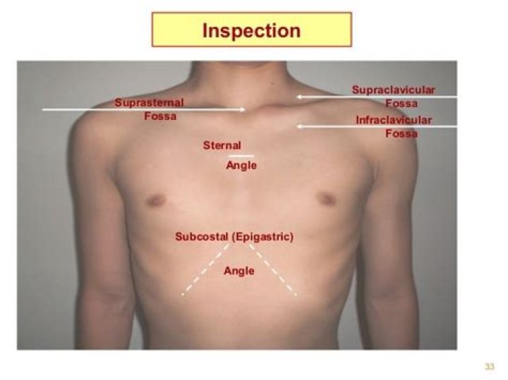
The word comes from jugum, Latin for yoke, presumably because yokes are carried on the neck. The jugular notch is the depression on the superior aspect of the manubrium at the hollow of the neck; it is also called the suprasternal notch. How do intercostal spaces feel?
From the angle of Louis, move your fingers to the right and you will feel a gap between the ribs. This gap is the 2nd Intercostal space. From this position, run your fingers downward across the next rib, and the next one. The space you are in is the 4th intercostal space. Where does the sternal angle lie?
The sternal angle is the angle formed between the manubrium of the sternum and the body of the sternum (manubriosternal junction), and is an important anatomical landmark. It marks the level of the 2nd pair of costal cartilages which lies at the level of the intervertebral disc between thoracic vertebrae 4 and 5. Why is rib 2 Atypical?
The first rib is atypical because it is wide and short, has two costal grooves, and one articular facet. The second rib is thin, long, and has a tuberosity on its superior surface for the attachment of the serratus anterior muscle. The eleventh and twelfth ribs have only one articular facet with no neck. What is a normal JVP height?
Normal: JVP is 6 to 8 cm above the right atrium. What is the sternal angle?
The sternal angle (also known as the angle of Louis or manubriosternal junction) is the synarthrotic joint formed by the articulation of the manubrium and the body of the sternum. The sternal angle is a palpable clinical landmark in surface anatomy. Why JVP is measured in cm of water?
JVP is a measure of Right atrial pressure. The number of cm of blood that rises as a blood column in the IJV is the pressure of the RA in cm of water. Explaination- Considering density of blood and water to be the same, cm of water is the same as saying cm of blood. How much is the sternal angle?
The sternal angle, which varies around 162 degrees in males, marks the approximate level of the 2nd pair of costal cartilages, which attach to the second ribs, and the level of the intervertebral disc between T4 and T5. In clinical applications, the sternal angle can be palpated at the T4 vertebral level. What is the importance of sternal angle?
The sternal angle is an important clinical landmark for identifying many other anatomical points: It marks the point at which the costal cartilages of rib two articulate with the sternum this is particularly useful when counting ribs to identify landmarks as rib one is often impalpable. Is it normal to see jugular vein pulsation?
If you ever see someone with a bulging neck vein, you're looking at the external jugular vein. When the jugular vein is visible, it's known as jugular vein distention (JVD). JVD is a sign of increased central venous pressure (CVP). That's a measurement of the pressure inside the vena cava. Why JVP is measured at 45 degrees?
Typically, this means that the venous waves are visible just above the clavicle when the patient is sitting at 30-45 degrees. With the JVP, the vessel is the internal jugular vein, and the fluid is the venous blood it contains. Look carefully on both sides of the neck for the JVP. What are examples of Synarthrosis joints?
A synarthrosis is a joint that is essentially immobile. This type of joint provides for a strong connection between the adjacent bones, which serves to protect internal structures such as the brain or heart. Examples include the fibrous joints of the skull sutures and the cartilaginous manubriosternal joint. Where is the Costochondral joint?
The costochondral joints are the joints between the ribs and costal cartilage in the front of the rib cage. They are hyaline cartilaginous joints (i.e. synchondrosis or primary cartilagenous joint). Costochondral joints articulate the lateral end of costal cartilage with eternal end of rib. Where is the Costosternal joint?
The sternocostal joints also known as sternochondral joints (or costosternal articulations), are synovial plane joints of the costal cartilages of the true ribs with the sternum, with the exception of the first, which is a synchondrosis since the cartilage is directly united with the sternum. What is a Synchondrosis joint?
Anatomical terminology. Where the connecting medium is hyaline cartilage, a cartilaginous joint is termed a synchondrosis. An example of a synchondrosis joint is the first sternocostal joint (where the first rib meets the manubrium). In this example, the rib articulates with the manubrium via the costal cartilage. What type of joint is Costochondral?
The costochondral joints are the joints between the ribs and costal cartilage in the front of the rib cage. They are hyaline cartilaginous joints (i.e. synchondrosis or primary cartilagenous joint). Each rib has a depression shaped like a cup that the costal cartilage articulates with. What is the Sternomanubrial joint?
The manubriosternal joint, sometimes referred to as the sternomanubrial joint, is the articulation between the upper two parts of the sternum, the manubrium and sternal body. Where is the Manubriosternal joint?
The manubriosternal joint is a type of secondary cartilaginous joint or symphysis, formed by the inferior border of the manubrium and the superior border of the sternal body. Both sides of the joint are irregular and undulating and covered with hyaline cartilage 2. What type of joint is the xiphoid process?
Anatomical terminology The xiphisternal joint (or xiphisternal synchondrosis) is a location near the bottom of the sternum, where the body of the sternum and the xiphoid process meet. It is structurally classified as a synchondrosis, and functionally classified as a synarthrosis. What is Sternocostal joint?
The sternocostal joints also known as sternochondral joints (or costosternal articulations), are synovial plane joints of the costal cartilages of the true ribs with the sternum, with the exception of the first, which is a synchondrosis since the cartilage is directly united with the sternum.